Chapter 1: The Brain in Your Belly (and Why That Phrase Is Misleading)
Summary
Your gut has its own nervous system, the enteric nervous system, built into the wall of the digestive tract. It contains hundreds of millions of neurons and can run digestive reflexes, such as the wave of squeezing that pushes food along, without any command from the brain. This chapter tours the gut wall, the two nerve networks inside it, the pacemaker cells that set its rhythm, and what happens when part of the network never forms (Hirschsprung disease). It also explains why the popular nickname "second brain" is useful but misleading, and it introduces the two tools you will use in every chapter: the Hype Meter and the six questions for reading a gut-brain claim.
Concepts Covered
- Digestive Tract
- Gut Wall Layers
- Smooth Muscle
- Central Nervous System
- Peripheral Nervous System
- Autonomic Nervous System
- Gut-Brain Axis
- Enteric Nervous System
- Myenteric Plexus
- Submucosal Plexus
- Enteric Neuron
- Enteric Glia
- Interstitial Cells of Cajal
- Peristalsis
- Peristaltic Reflex
- Migrating Motor Complex
- ENS Neuron Count
- Neural Crest
- Hirschsprung Disease
- Second Brain Metaphor
- Hype Meter
- Evidence Grade
Prerequisites
None. Just one year of high school biology. If you want a refresher on neurons, see From Neuron to Bedside.
Hi, I'm Vee, your vagus nerve guide
 I'm a vagus nerve fiber, the long line that carries messages between your gut and your brainstem, so I'll carry this book's messages too. You'll see me when there's a key idea to think about, a tip, a common mistake to dodge, a hard part worth pushing through, and a win to celebrate. Almost every gut-brain claim you'll see online starts with "your gut has its own brain," and you're about to find out how true that is. Let's check what the gut is really telling the brain!
1.1 Does Riya's Gut Have a Mind of Its Own?
Riya was 14 when she was diagnosed with Crohn's disease, after months of belly pain, diarrhea, weight loss, and a tiredness that sleep never fixed. On her first day back at Lakeview High, a friend asked what was wrong, and Riya, who did not feel like explaining inflammation in the hallway, said, "My gut has a mind of its own." Her friend laughed. Riya did too. But later, waiting for her first appointment with her pediatric gastroenterologist, Dr. Alvarez, she wondered whether the joke was closer to the truth than she thought. Does the gut actually make its own decisions? (Riya is a fictional student who appears throughout this book.)
In 1755, the Swiss scientist Albrecht von Haller noticed that a piece of intestine removed from an animal's body kept making its wave-like movements, even though it was no longer connected to the brain. Scientists still see the same thing today: cut every nerve to the brain and spinal cord, and a segment of intestine keeps pushing its contents along. So the gut really does run some of its own business. Whether that counts as a "mind" is what this chapter sorts out.
Riya's best friend Sam has his own version of the question. Sam runs cross-country, and before big races his stomach flips and churns. Is that his gut acting alone, or his brain talking to his gut? We'll start on that in Chapter 2. For now, remember two facts: the gut has its own local network, and it is also wired to the brain.
If you're going through this
If you have a gut condition, you have probably had to explain your body to people at bad moments, like Riya in that hallway. You don't owe anyone a full medical explanation. A short line such as "I have a health condition and might need the restroom" is enough, and your care team or school nurse can help you set up a plan with teachers. Learning how your gut works won't fix everything, but many people find it makes their body feel less mysterious and easier to talk about.
1.2 The Digestive Tract: A Muscular Tube With Layers
Before we meet the gut's neurons, we need to know what they live in. Picture a long garden hose that runs through your body. Now imagine that the hose is not plastic but made of several sleeves stacked inside each other, and that one of those sleeves is muscle that can squeeze. That is a good first picture of your digestive tract.
The digestive tract is the continuous tube that food travels through, from the mouth through the throat, esophagus, stomach, small intestine, and large intestine (colon) to the anus. Doctors also call it the gastrointestinal (GI) tract. In a living adult it is about 7.6 meters (25 feet) long, coiled up inside the abdomen, according to the open textbook Anatomy and Physiology 2e from OpenStax.
Along most of its length, the wall of the tube has the same basic structure. The gut wall layers are four tissue layers stacked from the inside (the open space where food travels, called the lumen) to the outside:
- Mucosa. The inner lining that touches food, with cells that absorb nutrients, make mucus, and release hormones, plus immune tissue.
- Submucosa. Loose connective tissue with blood vessels, lymph vessels, and glands. One of the gut's two nerve networks lives here.
- Muscularis. The muscle layer that squeezes. In the small intestine it has an inner layer of muscle wrapped in rings (circular muscle) and an outer layer running the long way (longitudinal muscle). The second nerve network sits between them.
- Serosa. A thin, slippery outer covering for the parts of the tract inside the abdomen.
The table below summarizes the four layers and adds which of the gut's two nerve networks each one holds. (We'll define those networks in Section 1.4.)
| Layer (inside to outside) | What it's made of | What it mainly does | Nerve network found here |
|---|---|---|---|
| Mucosa | Lining cells, mucus cells, hormone cells, immune tissue | Absorbs, secretes, senses what is in the gut | None of its own (nerve endings reach it) |
| Submucosa | Connective tissue, blood and lymph vessels | Supports the lining, carries blood | Submucosal plexus |
| Muscularis | Two sheets of smooth muscle | Mixes and moves contents | Myenteric plexus (between the sheets) |
| Serosa | Thin connective tissue covering | Lets the gut slide against nearby organs | None |
The muscle in the muscularis is a special kind. Smooth muscle is muscle made of small, spindle-shaped cells that contract slowly and can stay partly contracted for a long time, without you deciding to move it. You can choose to bend your elbow, but you can't choose to squeeze your small intestine. (A few spots, such as the mouth, throat, upper esophagus, and the outer ring of muscle at the anus, use skeletal muscle instead, which is why you control swallowing.) When the circular muscle contracts, it pinches the tube narrower, like a hand closing around a toothpaste tube.
Coordinate that squeeze into a traveling wave, and you can push food a long way. The rest of this chapter is about who does the coordinating.
1.3 Two Nervous Systems, One Body
You already know the basic layout of the nervous system from biology class, so here is a quick refresher. Neurons are cells that carry electrical signals and pass messages to other cells at junctions called synapses. The central nervous system (CNS) is the brain and spinal cord, the body's main processing center. The peripheral nervous system (PNS) is everything outside the brain and spinal cord: the nerves that branch out to your skin, muscles, and organs, and the clusters of nerve cell bodies (ganglia) along the way. For a deeper review of neurons and brain anatomy, see From Neuron to Bedside, Chapter 1 and Chapter 4.
Part of the peripheral nervous system runs the body's background jobs, the ones you never think about, like heart rate, sweating, and digestion. The autonomic nervous system is this automatic, involuntary part of the nervous system that controls internal organs, glands, and smooth muscle. Textbooks usually describe two branches:
- The sympathetic branch, often called "fight or flight." When it is active, it tends to slow digestion: secretions drop and gut muscle movement is held back.
- The parasympathetic branch, often called "rest and digest." When it is active, it tends to speed digestion up. The vagus nerve (Vee's home) is its biggest gut connection.
Early in the 1900s, the British physiologist John Newport Langley proposed that the autonomic nervous system actually has three divisions: sympathetic, parasympathetic, and a third one inside the gut wall. Many reviews still describe it that way, and that third division is the star of this chapter.
Think of a city. The brain and spinal cord are city hall. The sympathetic and parasympathetic nerves are phone lines to each neighborhood. And the gut has its own neighborhood council that handles local problems, like clearing a blocked street, without calling city hall. City hall can call in and change priorities, and the council sends reports back, but routine work needs no permission.
All of these pieces talk to each other. The gut-brain axis is the two-way communication system linking the gut (its nervous system, hormones, immune cells, and microbes) with the brain. Signals travel both up and down, and nerves are only one route; later chapters cover hormones, immune messengers, and chemicals made by gut microbes. This chapter is about the local network at the gut end of the conversation.
1.4 Meet the Enteric Nervous System
The enteric nervous system (ENS) is the network of neurons and support cells built into the wall of the digestive tract, running from the esophagus to the anus. ("Enteric" comes from a Greek word for intestine.) A 2020 review by Spencer and Hu describes the gut as the only internal organ that evolved its own complete nervous system, one that can work fully independently of input from the brain and spinal cord.
Imagine two layers of fishing net wrapped around the garden hose from Section 1.2, one tucked under the lining and one sandwiched inside the muscle. Each knot is a small cluster of nerve cell bodies called a ganglion, and the strings are bundles of nerve fibers.
The myenteric plexus (also called Auerbach's plexus) is the outer network of ganglia, located between the circular and longitudinal muscle layers. It runs the whole length of the gut and mainly controls movement: the rhythm and strength of contractions. ("Plexus" means a network of nerves.)
The submucosal plexus (also called Meissner's plexus) is the inner network of ganglia, located in the submucosa just under the lining. It is found mainly in the small and large intestines. It mostly controls what the lining does, such as releasing fluid and mucus, and it helps sense what is in the gut.
So many new names
 I know, "myenteric" and "submucosal" look like a lot on day one. Here's my trick: myenteric sits in the muscle and runs movement, while submucosal sits sub (under) the mucosa and runs the lining.
An enteric neuron is a nerve cell whose cell body sits in the gut wall, inside one of the ENS ganglia. A 2020 review by Fleming and colleagues says the ENS contains up to 20 types of neurons, in three big job categories:
- Sensory neurons (scientists call them intrinsic primary afferent neurons) detect stretch of the gut wall and chemicals in the gut.
- Interneurons pass signals up and down the gut, connecting sensory neurons to motor neurons.
- Motor neurons send signals to muscle and glands. Some are excitatory and make muscle contract, often using the chemical messenger acetylcholine. Others are inhibitory and make muscle relax, often using nitric oxide.
That is everything you need to build a reflex, all inside the gut wall. Hold on to that idea for Section 1.7.
The second kind of cell is the support crew. Enteric glia are non-neuron cells in the ENS that surround and support enteric neurons and help regulate gut function. Scientists once thought glia were just packing material, but a 2021 review by Seguella and Gulbransen says that view has been refuted. Enteric glia appear to help regulate gut reflexes, the gut lining's barrier, and immune responses, though much of that evidence comes from animals. They also outnumber enteric neurons; one review estimates roughly 2 to 3 glia per neuron, a rough figure from a single source.
1.5 How Many Neurons Are in Your Gut?
This is the number wellness posts love to quote. The ENS neuron count is the estimated total number of neurons in the human enteric nervous system. The careful version of the fact, used throughout this book, is this:
The enteric nervous system contains hundreds of millions of neurons, about 400 to 600 million by common estimates, roughly as many as the spinal cord.
Why "estimates"? Nobody can count every neuron in a gut several meters long, so scientists count small samples and multiply up, and different methods give different answers. A 2016 review by Rao and Gershon says "more than 100 million," a 2019 review by Schneider, Wright, and Heuckeroth says "more than 500 million," and the 2020 Fleming review says "approximately 400 to 600 million." Some textbooks, including OpenStax, give about 100 million. The order of magnitude is solid; the exact figure is not.
Now compare that with the brain. In 2009, a team including Frederico Azevedo and Suzana Herculano-Houzel counted cells in adult male human brains using a cell-counting method called the isotropic fractionator. They found about 86 billion neurons on average. Here is the comparison side by side:
| Structure | Approximate number of neurons | Source |
|---|---|---|
| Enteric nervous system (gut wall) | About 400 to 600 million (estimates vary) | Fleming 2020; Schneider 2019; Rao and Gershon 2016 |
| Spinal cord | Roughly similar to the ENS | Fleming 2020; Rao and Gershon 2016 |
| Brain | About 86 billion | Azevedo et al. 2009 |
Do the math: 86 billion divided by 500 million is about 170, so the brain has well over a hundred times more neurons. Still, Rao and Gershon note that the ENS has more neurons than all the body's other peripheral ganglia combined, and far more than the number of vagus nerve fibers carrying commands down to the gut. The brain doesn't have enough wires to micromanage the gut, so the gut has to handle much of its own control.
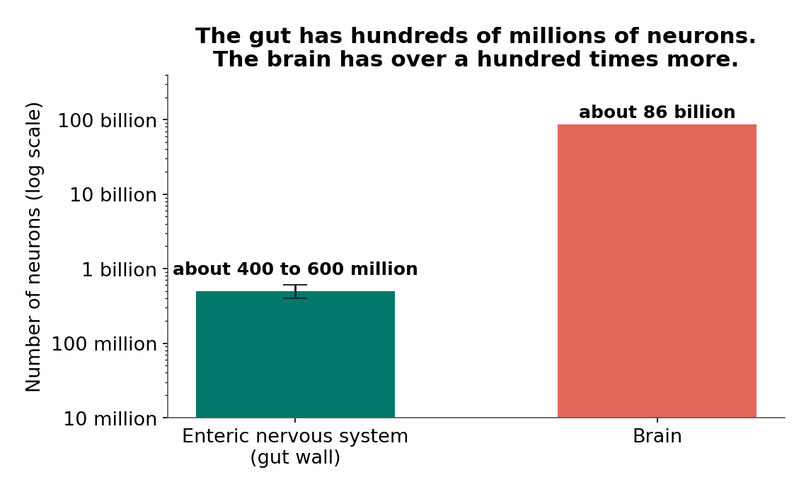
Source: Fleming et al. 2020; Schneider et al. 2019; Azevedo et al. 2009. The scale is logarithmic: each step up is ten times bigger.
Big for a nerve net, small for a brain
 When you see "your gut has millions of neurons," both parts can be true: it's a huge number for a nerve network, and a tiny one next to your brain's 86 billion. Always ask, "Compared with what?"
1.6 The Gut's Pacemakers: Interstitial Cells of Cajal
Your heart has pacemaker cells that set its beat. Your gut has something similar. Think of a drummer in a marching band: the drummer doesn't decide where the band marches, but the beat tells everyone when to step.
Interstitial cells of Cajal (ICC) are specialized cells in the gut's muscle layers that generate rhythmic electrical signals, called slow waves, which set the timing of smooth muscle contractions. They are named after Santiago Ramón y Cajal, a Spanish scientist and Nobel Prize winner famous for his studies of nerve cells. ICC are neither neurons nor muscle cells, but they are electrically connected to the muscle.
A slow wave is a gentle, repeating rise and fall in the electrical charge of muscle cells that spreads from the ICC and sets the timing of contractions. ICC also help pass signals from enteric motor neurons to the muscle, so how strongly the muscle squeezes depends on the neurons too. ICC set the beat; the enteric neurons decide which steps happen.
How do scientists know ICC are the pacemakers? ICC carry a receptor protein called Kit on their surface. In 1994, Ward, Sanders, and colleagues studied mice with mutations in the gene for Kit. These mice had very few ICC in the myenteric region of the small intestine, and recordings from their intestinal muscle showed that the electrical slow waves were missing. Normal sibling mice always had slow waves, and a 1995 study by Huizinga and colleagues found the same link. The mutant muscles still responded when enteric nerves were stimulated, so nerve control can develop without ICC.
Notice the kind of evidence. These were mouse studies, but they were experiments that tested cause: change one gene, lose the cells, lose the rhythm. Human disease data later supported the same picture, so "ICC are gut pacemakers" rates as Strong.
There is a catch. The Spencer and Hu review notes that in some experimental animals, the small intestine's pacemaker ICC can be removed without wrecking normal function, while mice that lack an ENS cannot push food along and die. The beat helps, but the band can't march without the enteric neurons.
1.7 Peristalsis: How Food Gets Pushed
Squeeze the bottom of a toothpaste tube and slide your fingers toward the cap. Now imagine the tube could do that by itself, sensing where the paste is and squeezing just behind it. That's what your intestine does.
Peristalsis is a wave of muscle contraction that travels along the digestive tract and pushes its contents forward, toward the anus. (The intestine also makes mixing movements, but peristalsis is how food makes forward progress.)
What triggers each wave? The peristaltic reflex is a local reflex of the enteric nervous system in which stretching of the gut wall causes the muscle behind the stretched spot (on the mouth side) to contract and the muscle ahead of it (on the anus side) to relax. In 1899, the physiologists Bayliss and Starling, working with loops of dog intestine, summarized it as a "law of the intestine": local stimulation produces excitation above the stimulated spot and inhibition below it. During World War I, the scientist Paul Trendelenburg studied isolated segments of intestine, completely cut off from the brain and spinal cord, and gave the response the name we still use today: the peristaltic reflex.
Here is the reflex step by step, using the three neuron types from Section 1.4:
- Stretch. A ball of food (doctors call it a bolus) stretches the gut wall.
- Sense. Stretch-sensing enteric neurons in that spot fire.
- Relay. Interneurons carry the signal in two directions: back toward the mouth side and forward toward the anus side.
- Squeeze behind. On the mouth side, excitatory motor neurons tell the circular muscle to contract. The tube pinches closed behind the bolus.
- Relax ahead. On the anus side, inhibitory motor neurons tell the circular muscle to relax. The tube opens up in front of the bolus.
- Repeat. The bolus moves into the relaxed section, stretches a new spot, and the cycle starts again a little farther along.
Step 5 is as important as step 4: if the tube ahead stays clamped shut, squeezing behind the food just builds pressure. That is exactly what goes wrong in Hirschsprung disease. And notice what is not on the list: the brain. It can speed the process up or slow it down, but the reflex itself is local.
One correction to an older idea: many textbooks said serotonin released from the gut lining triggers peristalsis. The Spencer and Hu review says evidence, mostly from animals, now suggests it is not required, although it can change how the waves behave. (More on gut serotonin in Chapter 5.)
In the simulation below, "stretch sensitivity" means how easily the stretch-sensing neurons fire, the red zone is contracting circular muscle behind the bolus, and the green zone is relaxing muscle ahead of it.
Diagram: Peristalsis Builder
Open the Peristalsis Builder fullscreen
MicroSim specification: Peristalsis Builder
Type: microsim
sim-id: ch01-peristalsis-builder
Library: p5.js
Status: Implemented
Bloom Level: Understand
Bloom Verb: explain
Learning Objective: The learner will explain how the peristaltic reflex moves a bolus as a local enteric circuit (stretch, then contraction behind and relaxation ahead) and predict why the bolus stalls where enteric neurons are missing.
Canvas: 400 px drawing area plus 120 px controls (iframe 522 px); responsive to about 380 px wide.
Controls: Start/Pause, Reset, Show labels; ENS select (Working everywhere; Missing at the end, like Hirschsprung; Switched off everywhere); Stretch sensitivity slider (1 to 10, step 1, default 6, arbitrary units).
Rules: The reflex fires when the stretch signal (5 x sensitivity / 6) is >= 3.5, so sensitivity 1 to 4 gives no reflex and 5 to 10 does; speed rises with sensitivity. The bolus advances only if the segment ahead has enteric neurons and can relax. A no-neuron segment stays narrowed (contracted). With no ENS, pacemaker ripples continue but do not move the bolus. Material backs up behind a blockage.
Feedback: A status panel names the state (paused, firing, below trigger, stalled, no ENS, delivered) and shows deliveries and last trip time (illustrative seconds).
Provenance: Section 1.7 (Spencer and Hu 2020; Bayliss and Starling 1899); contracted no-neuron segment (Heuckeroth 2018). All numbers are illustrative.
Start with the ENS working everywhere and press Start. Watch the red "squeeze" zone chase the bolus while the green "relax" zone opens the way ahead; the gold neuron dots light up only near the food. Then drag the stretch sensitivity down to 4 or lower. The signal falls below the trigger level, so the reflex never fires. Raise it again and compare trip times at 6 and 10.
Now predict: if the last part of the tube has no enteric neurons, where will the food stop? Choose Missing at the end (like Hirschsprung) and test it. The bolus stalls at the edge of the no-neuron zone, which stays squeezed because it has no inhibitory neurons, and the tube behind it swells. Finally, try Switched off everywhere: the pacemaker ripples continue, but they can't move anything.
What this model leaves out
- A real gut is several meters long with many boluses, mixing movements, and fluid, not one ball in a straight tube.
- Real reflexes travel through chains of interneurons and dozens of neuron types; the sim shows only "sense, squeeze, relax."
- The brain, spinal cord, and vagus nerve can speed up or slow down peristalsis; the sim leaves out all outside nerve input and hormones.
- Speeds, stretch values, and the trigger level are made-up teaching numbers, not measurements.
- In real Hirschsprung disease, the length of the no-neuron segment varies from child to child, and the swelling builds over days, not seconds.
1.8 Between Meals: The Migrating Motor Complex
What does the gut do when you haven't eaten for a while? It cleans house.
The migrating motor complex (MMC) is a repeating pattern of strong contractions that sweeps slowly along the stomach and small intestine during fasting, between meals. Picture a street sweeper that comes through on a schedule when there's no traffic, pushing leftover debris onward toward the large intestine.
A 2012 review by Deloose and colleagues describes the MMC as a cyclic pattern that occurs during fasting and is interrupted by eating. It is divided into phases, and the most active phase (phase III) is a burst of strong contractions that starts in the lower stomach or upper small intestine and travels downstream. In humans, phase III contractions come back roughly every 90 to 120 minutes, according to a 2012 review by Toku Takahashi. Because problems with the MMC have been linked to an overgrowth of bacteria in the small intestine, older papers nicknamed it the gut's "housekeeper."
The MMC also gives us more evidence for the ENS's independence. The Deloose review notes that cutting the vagus nerve (a surgery called vagotomy) stops the MMC's motor activity in the stomach but leaves the repeating activity in the small intestine intact.
Hormones help control the MMC too: one gut hormone, motilin, rises and falls with its phases, and some recent research suggests these contractions may help signal hunger, an idea still being tested. The key point: even with no food to push, the ENS keeps its own timetable.
1.9 Where Enteric Neurons Come From: The Neural Crest
Every neuron in your ENS started somewhere else. Early in development, the embryo forms a tube of tissue that will become the brain and spinal cord. Along the edges of that tube, a special group of cells breaks away and travels through the body. The neural crest is this group of embryonic cells that forms at the edge of the developing neural tube and migrates throughout the embryo, giving rise to many cell types, including the neurons and glia of the enteric nervous system.
Nearly all enteric neurons and glia come from the neural crest, and most come from one region of it, called the vagal neural crest, located in the lower part of the developing hindbrain, near the junction between brain and spinal cord. (A smaller group from the lower end of the neural tube, the sacral neural crest, adds cells mainly to the end of the colon.) A 2017 review by Nagy and Goldstein describes how these cells enter the top of the gut in about the fourth week of development in humans and then migrate down the gut tube from top to bottom, colonizing the entire length of the gut. In bird and rodent embryos, the leading edge of these migrating cells moves at about 40 micrometers per hour.
Think of it like settlers spreading across a continent from one coast: the last region to be reached is the far end. That's why, if the migration falls short, the part left without neurons is almost always the end of the bowel.
The trip depends on chemical signals. Nagy and Goldstein call RET, a receptor protein on the migrating cells, critically important for ENS development, and changes in the RET gene and several others raise the chance that migration falls short.
1.10 When the Network Is Missing: Hirschsprung Disease
Hirschsprung disease is a condition present from birth in which enteric neurons are missing from the last part of the large intestine, so that segment cannot relax and push stool forward, causing a blockage. A 2018 review by Heuckeroth puts it plainly: when the ENS is missing, there are no nerve-driven pushing movements, and the bowel stays contracted. Without neurons, the gut doesn't go limp, as many people guess. It stays squeezed, because the inhibitory neurons that would tell it to relax aren't there. Stool backs up behind it, just like in the sim's "missing at the end" setting.
The no-neuron stretch always starts at the rectum, but its length varies: NIDDK says that in about 80% of children it is short (the rectum and sometimes the lowest part of the colon), while in about 5% to 10% nerve cells are missing from the whole large intestine, and sometimes part of the small intestine too.
According to the National Institute of Diabetes and Digestive and Kidney Diseases (NIDDK), about 1 in 5,000 newborns has Hirschsprung disease. The condition is named after Harald Hirschsprung, a Danish children's doctor who presented two cases at a medical conference in Berlin in 1886 and published his description in 1888. Some babies show signs of a blocked intestine soon after birth, such as not passing a first stool within 48 hours, green or brown vomit, and a swollen belly. Other children are diagnosed later because of long-lasting constipation that doesn't get better with usual treatments.
Hirschsprung disease is clear human evidence that the ENS is not optional. These children have a working brain, spinal cord, and vagus nerves; missing the local network in one segment is enough to stop the bowel.
For families
If a baby or child in your family has Hirschsprung disease, it helps to know where it comes from: nerve cells in the gut didn't finish developing before birth, and certain genes raise the chance. Many parents wonder whether they caused it, and that is a good question to bring to the care team, who can explain what is known for your child. After surgery, many children do well, but some have ongoing issues such as constipation or trouble with bowel control, so regular follow-up with the care team matters. Siblings may feel left out during hospital stays; a simple explanation like "the end of the baby's gut didn't get its nerve cells, and the doctors fixed the part that wasn't working" can help them feel included.
1.11 "Second Brain": A Useful Nickname, a Misleading Claim
By now you can see why people call the ENS a "brain in your belly": it is large, has its own sensory and motor circuits, and can run without the brain. In 1998, the Columbia University scientist Michael D. Gershon published a popular book called The Second Brain, and the nickname spread. Researchers still use it. A 2019 scientific review is titled "Unexpected roles for the second brain."
The second brain metaphor is the comparison of the enteric nervous system to a brain, which highlights that it is large, complex, and able to work independently, but which can mislead people into thinking the gut thinks, feels, or decides like the brain does. Here is where the comparison holds up and where it breaks down:
| What the ENS does (supported) | What the "second brain" phrase can wrongly suggest (not supported) |
|---|---|
| Senses stretch and chemicals in the gut | That the gut has thoughts or feelings |
| Runs local reflexes like peristalsis | That the gut makes decisions or plans |
| Controls secretion, mixing, and blood flow in the gut wall | That "gut instinct" comes from gut neurons thinking on their own |
| Works without input from the brain | That the ENS is equal to the brain |
| Sends signals to the brain and receives signals from it | That the gut has more neurons than the brain |
None of the research reviews used for this chapter describe the ENS as thinking, reasoning, or feeling. They describe movement, secretion, blood flow, and barrier and immune roles. A sophisticated reflex network is impressive enough without exaggeration.
So what about "gut feelings," like Sam's pre-race butterflies? Those feelings are real, but you feel them with your brain. Signals from the gut travel up to the brain, mostly through the vagus nerve and spinal nerves, and the brain turns them into sensations like nausea, fullness, or that fluttery feeling. The brain also sends signals back down that change how the gut behaves. That two-way traffic is the subject of Chapters 2 and 7. The gut senses and reports; it doesn't think.
Watch the word "thinks"
 The structure fact ("your gut has its own nervous system") is rock solid, but the leap to "your gut thinks and feels" is not. When a post slides from the first claim to the second, that slide is exactly where hype sneaks in.
1.12 The Hype Meter: Grading Gut-Brain Claims
The gut-brain axis is real science, and also one of the most oversold topics in wellness. A finding in a few mice can become "this supplement cures anxiety" by the time it reaches your feed.
The Hype Meter is this book's four-level scale for grading how well the evidence supports a popular gut-brain claim, from Strong to Moderate to Preliminary to Hype. When we apply it, we give a claim an evidence grade, the rating that summarizes how strong and how direct the scientific support for a specific claim is, as it is actually stated.
Strong means consistent evidence in people, usually from large or repeated studies, randomized trials (experiments where people are assigned to groups by chance), or well-established physiology. Moderate means good animal evidence plus some human evidence, or a strong, repeated link in people whose cause is still unclear. Preliminary means small, early, short, or mostly animal studies. Hype means a popular claim the evidence does not support, or one that stretches a real finding far beyond what it shows. The table below summarizes the four grades.
| Grade | Typical evidence | What it means for you |
|---|---|---|
| Strong | Repeated human studies, trials, textbook physiology | Treat as established (still not personal medical advice) |
| Moderate | Animals plus some humans, or a solid link without a proven cause | Probably real in part; details still being worked out |
| Preliminary | Few, small, short, or animal-only studies | Worth watching; don't act on it yet |
| Hype | Popular claim not backed by evidence | Be skeptical, especially if someone is selling something |
Two rules make the Hype Meter work. First, grade the claim as stated, not the topic. "Your gut has its own nervous system" and "your gut thinks" start from the same fact but get very different grades. Second, grades can change as new studies come out; this book logs changes on its Accuracy page. And a "Strong" grade for a treatment never means it is right for you personally. That is a conversation for you and your doctor. Here are three claims you're likely to hear about this chapter's topic.
Hype Meter: "Your gut can move food without your brain"
Hype Meter: "Your gut is a second brain that thinks and feels"
Hype Meter: "Your gut has more neurons than your brain"
1.13 How to Read a Gut-Brain Claim: Six Questions
A grade is only as good as the reasoning behind it, so every chapter applies the same six questions to at least one real study. You can use them on any health headline, ad, or post.
- Mouse or human? Many gut-brain findings come from mice, including mice raised with no microbes at all. Mouse results often don't carry over to people.
- Correlation or cause? Two things happening together is a correlation, not proof that one causes the other. The arrow might point the other way, or a third factor might explain both. Showing cause usually takes an experiment that changes one thing on purpose and compares with a group where nothing changed.
- How many? Small studies swing wildly by chance. A result in 12 people or 8 mice is a hint, not an answer.
- How long? A two-week change might not last, and a long-term harm might not show up yet.
- Who paid for it? Funding doesn't make a study wrong, but it's worth knowing who benefits. Good papers include a funding and conflict-of-interest statement.
- Is something being sold? If a claim ends with a product, a test kit, a diet plan, or a program, read it twice as carefully.
Use the questions on good studies too
 These questions aren't only for spotting bad science. Use them on excellent studies too, because even a great study answers only the question it actually asked.
Let's practice on a real, high-quality study from this chapter's topic. In 2020, a team at the Broad Institute of MIT and Harvard (first author Drokhlyansky, with senior authors Regev and Xavier) published an atlas of enteric neurons in the journal Cell. They developed new methods to read which genes are active in single cells. In mice, they profiled 1,187,535 cell nuclei, which included 5,068 neurons from the small intestine and colon. In humans, they profiled 436,202 nuclei from colon tissue and recovered 1,445 neurons. The human tissue came from the healthy-looking edges of colon sections removed during surgery from 16 people with colon cancer, aged 35 to 90. The team found dozens of distinct types of enteric neurons, including 21 neuron subsets in one mouse dataset.
How to read a gut-brain claim: the 2020 enteric neuron atlas
| Question | This study |
|---|---|
| Mouse or human? | Both. Most of the data came from mice (including 29 mice for one detailed dataset), plus colon tissue from 16 human patients. |
| Correlation or cause? | Neither, really. This is a descriptive map: it catalogs which genes each neuron type uses. It can suggest what neurons might do, but it doesn't test cause. |
| How many? | Very large numbers of cells (over 1.6 million nuclei) but only 1,445 human neurons from 16 people, all of whom had colon cancer surgery. |
| How long? | A snapshot. Each sample was taken once, so it can't show changes in one person over time (though the mouse data compared ages and times of day). |
| Who paid for it? | The paper lists the Klarman Cell Observatory, the Howard Hughes Medical Institute, several NIH grants, the Manton Foundation, the Helmsley Charitable Trust, the Crohn's and Colitis Foundation, and others. |
| Is something being sold? | No product is sold to the public. However, several authors are co-inventors on a patent application covering the methods, and some hold equity in or work for biotech or drug companies, as the paper's conflict-of-interest statement discloses. |
The conclusion: this is careful, valuable research supporting the idea that the ENS has many neuron types. It does not show that any neuron type causes a disease, and the human tissue came from adults with cancer, who might not represent healthy teens. The patent and company ties don't make the findings wrong; they are facts a careful reader notes.
At the Bedside: A Newborn Who Can't Pass Stool
Meet the patient (fictional)
Mateo is a 2-day-old baby in a hospital newborn nursery. His parents notice that he hasn't had his first bowel movement, even though other babies born the same day have. By his second day, his belly looks round and tight, he isn't feeding well, and he vomits green fluid.
What the doctor checks: The newborn team takes a family history, examines Mateo's swollen belly, and notes that he has not passed a first stool within 48 hours of birth, a key warning sign. Imaging, such as a contrast enema (a liquid that shows up on X-rays, put in through the rectum), shows the shape of the large intestine. The test that confirms the diagnosis is a rectal biopsy: tiny pieces of rectal tissue examined under a microscope.
Connecting the signals: No stool plus a swollen belly means a blockage, but nothing is physically stuck. The pathologist finds no ganglion cells (the enteric neuron clusters) in the submucosal layer of the biopsy samples, the layer a rectal biopsy usually reaches. In Hirschsprung disease, the neurons are missing from both nerve networks in the affected segment. Without enteric neurons, the last part of his colon can't relax to let stool through.
Diagnosis: Hirschsprung disease.
Why it happens: Before Mateo was born, the neural crest cells that should have colonized his gut from top to bottom did not finish colonizing the end of his colon. The segment without enteric neurons stays contracted, so the peristaltic reflex in the healthy gut above it pushes stool into a section that never opens. That is the "missing at the end" setting of the Peristalsis Builder.
The mind-body side: Mateo can't explain how he feels, but his parents carry a lot of fear and guilt in the first days, and many ask whether they caused this. The care team explains what is known: the nerve cells in the gut didn't finish developing before birth, and genes play a role. Neonatal nurses, surgeons, and social workers may all help the family understand the surgery, hold and feed their baby when possible, and plan for life at home. As children grow up, bowel control problems can affect confidence at school, and care teams can connect families with support.
Treatment today: The main treatment is surgery called a pull-through procedure. The surgeon removes the part of the large intestine that is missing nerve cells and connects the healthy part to the anus. Some children first need ostomy surgery, which creates a usually temporary opening (a stoma) on the belly for stool to leave while the intestine heals. According to NIDDK, children most often feel better after surgery, but about half may have ongoing problems such as constipation, trouble with bowel control, or a serious gut inflammation called Hirschsprung-associated enterocolitis, which needs hospital care right away.
What's new in research: In 2016, a team led by Fattahi and Studer reported in Nature that they could grow early enteric nervous system cells from human stem cells in the lab. When they transplanted these cells into the colons of mice with a Hirschsprung-like condition, the cells spread into the gut and the mice were more likely to survive, although the researchers said the reason was unclear. Evidence grade: Preliminary. These are mouse and lab results; cell therapy for Hirschsprung disease is experimental and not a standard treatment. In 2025, a Massachusetts General Hospital team led by pediatric surgeon Allan Goldstein reported that its mouse studies of a therapy using a patient's own gut nerve stem cells were largely complete and that it planned to ask the FDA to begin a first trial in people. As of October 2026, we found no cell therapy trial for Hirschsprung disease registered on ClinicalTrials.gov, and no results in children have been reported.
Your turn: reason through a variation
A 4-year-old has had constipation for years that hasn't improved with the usual medicines, and his belly is often swollen. His parents read about Hirschsprung disease and ask, "Isn't that only in newborns?" How would you reason about this?
Answer: No. Many children with Hirschsprung disease show symptoms as newborns, but not all. NIDDK says about half are diagnosed in the first year of life and about 80% by age 7. Children diagnosed later often have a short segment without neurons and milder symptoms. Constipation that doesn't respond to usual treatment is one sign in older children. Many other things cause constipation, so only the care team can sort it out, and a rectal biopsy is still the test that confirms or rules out Hirschsprung disease.
This is a fictional teaching case. It is not medical advice. If you or someone you know has symptoms like these, talk to a doctor.
Common Misconceptions
Myth: The brain has to send a command for every movement of the gut.
Reality: The enteric nervous system runs the peristaltic reflex locally. Pieces of intestine cut off from the brain keep pushing their contents along. The brain adjusts gut activity but isn't needed for the basic reflex.
Myth: The gut has more neurons than the brain.
Reality: The ENS has about 400 to 600 million neurons by common estimates, roughly as many as the spinal cord. The brain has about 86 billion, well over a hundred times more.
Myth: "Second brain" means the gut is equal to the brain and can think or feel.
Reality: The ENS is a large, independent reflex network. It senses and controls movement, secretion, and blood flow in the gut. Sensations like butterflies or nausea are created by the brain from signals the gut sends up.
Myth: If part of the gut has no neurons, it just goes limp.
Reality: In Hirschsprung disease, the segment without enteric neurons stays contracted because there are no inhibitory neurons to relax it. That is why it blocks stool instead of letting it slide through.
Myth: The pacemaker cells of the gut can move food on their own.
Reality: Interstitial cells of Cajal set the rhythm of muscle activity, but animals without an enteric nervous system can't push food along, even when their pacemaker cells are present. The beat needs the neurons.
Your Gut Check
Test yourself. Try each one before you open the answer.
1. Claim Check: "Your gut has more neurons than your brain. That's why you should always trust your gut over your head!"
Grade: Hype. The first sentence is false: the ENS has about 400 to 600 million neurons, while the brain has about 86 billion. The second doesn't follow even from true facts: the ENS runs reflexes, not decisions, and "gut feelings" are sensations your brain creates from gut signals. A careful version would say: "Your gut has its own network of hundreds of millions of neurons that runs digestion locally and sends signals to your brain."
2. Predict: Riya's cousin had surgery years ago in which the vagus nerve to his stomach was cut. Riya asks Dr. Alvarez whether food can still move through his small intestine. What would you predict, and why?
Food should still move through his small intestine, because the peristaltic reflex is run by the ENS inside the gut wall. Research on the migrating motor complex also shows that after vagotomy, the stomach part of the between-meal pattern stops, but the small intestine's repeating activity continues. His stomach's movements might change, since the vagus nerve plays a bigger role there.
3. Explain: A newborn with Hirschsprung disease has a swollen belly. Use the peristaltic reflex to explain why the belly swells above the part of the colon without neurons.
In the healthy gut above the missing segment, stool stretches the wall, sensory enteric neurons fire, and the muscle squeezes behind the stool as it should. But the segment ahead has no inhibitory motor neurons, so it can't relax and stays contracted. Stool and gas keep getting pushed toward a section that never opens, so they pile up and stretch the gut above it, which makes the belly swell.
4. Apply: In a lab, a scientist places a small balloon inside an isolated segment of guinea pig intestine and inflates it. The segment has no connection to the brain or spinal cord. What will happen to the muscle on each side of the balloon?
The balloon stretches the gut wall, which triggers the peristaltic reflex. On the mouth side of the balloon, the circular muscle should contract; on the anus side, it should relax. This is Bayliss and Starling's "law of the intestine." It works because sensory neurons, interneurons, and motor neurons are all inside the gut wall, so no brain connection is needed.
5. Read the data. In the 2020 enteric neuron atlas (Drokhlyansky et al., Cell), researchers profiled cell nuclei from gut tissue and counted how many turned out to be neurons. Here are their numbers:
| Sample | Nuclei profiled | Neurons recovered |
|---|---|---|
| Mouse small intestine and colon | 1,187,535 | 5,068 |
| Human colon (16 patients) | 436,202 | 1,445 |
5. What does this data show, and what can't it show?
It shows that neurons are a tiny fraction of the cells in gut tissue: about \( 5{,}068 \div 1{,}187{,}535 \approx 0.4\% \) in the mouse samples and about \( 1{,}445 \div 436{,}202 \approx 0.3\% \) in the human samples. That's why the team needed special methods to find them. It can't give the total neuron count of a whole gut, because these were small samples processed to enrich and filter cells, not to count everything. It also can't show cause or say much about healthy teens, since the human tissue came from 16 adults having cancer surgery. A helpful follow-up: a similar atlas from a wider range of healthy donors, including younger people.
Exercises
Remember
- Name the four layers of the gut wall, from the inside out.
Answer
Mucosa, submucosa, muscularis, and serosa.
- Where is the myenteric plexus located, and what does it mainly control?
Answer
It sits between the circular and longitudinal muscle layers of the gut wall and mainly controls gut movement (the rhythm and strength of contractions).
Understand
- Explain why relaxation of the muscle ahead of a bolus is as important as contraction behind it.
Answer
Squeezing behind the bolus only moves it forward if the tube ahead opens. If the muscle ahead stays contracted, pressure builds but the bolus can't advance, which is what happens at the no-neuron segment in Hirschsprung disease.
- In your own words, explain the difference between the ENS neuron count being "solid in order of magnitude" and "uncertain in exact number."
Answer
Scientists agree the ENS has hundreds of millions of neurons, not thousands and not billions. But because they count small samples and scale up with different methods, estimates range from more than 100 million to about 600 million, so no single exact number is reliable.
Apply
-
A wellness video says, "Your gut is your second brain, so it knows what foods your body needs." Use the second brain metaphor table in Section 1.11 to identify which part of this claim is supported and which part is not, and give it an evidence grade.
-
Using the MicroSim, record the trip time at stretch sensitivity 4, 6, 8, and 10 (press Reset before each run; if the food never arrives, record "no trip"). Describe the pattern, then explain what "stretch sensitivity" represents in real enteric neurons and why the model's numbers can't be compared to a real gut.
Analyze
-
Compare the evidence from the 1994 Kit mutant mouse experiments with the evidence from children with Hirschsprung disease. For each, answer two of the six questions (mouse or human, and correlation or cause). Which tells you more about humans, and which is better at showing cause?
-
Riya's Crohn's disease affects the end of her small intestine and part of her colon, and her enteric nervous system is present. A newborn with Hirschsprung disease is missing enteric neurons in the end of the colon. Analyze how the cause of a bowel problem differs between "the nerve network is missing" and "the gut is inflamed," and why this difference matters for treatment.
Evaluate
-
Some scientists think the "second brain" nickname helps the public appreciate the ENS; others think it causes more confusion than it's worth. Write a one-paragraph judgment: should this textbook keep using the phrase? Support your position with at least two pieces of evidence from this chapter.
-
Evaluate this headline: "Scientists map 21 types of gut neurons, showing your gut is smarter than you think." Use at least four of the six questions and the claim-box in Section 1.13 to decide what grade the headline deserves.
Create
-
Design a simple experiment, using isolated segments of animal intestine, that would test whether a particular drug blocks the relaxation part of the peristaltic reflex but not the contraction part. Describe your groups, what you would measure, and the result that would support your hypothesis.
-
Create a short social-media post (under 280 characters) that correctly explains what the enteric nervous system does, uses the "second brain" phrase responsibly, and would earn a Strong grade on the Hype Meter.
Key Takeaways
- The digestive tract is a muscular tube with four wall layers; its smooth muscle squeezes without conscious control.
- The enteric nervous system, built into the gut wall, has two networks: the myenteric plexus (mainly movement) and the submucosal plexus (mainly the lining). It contains hundreds of millions of neurons, about 400 to 600 million by common estimates, roughly as many as the spinal cord, and far fewer than the brain's 86 billion.
- The peristaltic reflex is a local circuit: stretch triggers contraction behind the food and relaxation ahead of it, with no command from the brain needed. Between meals, the migrating motor complex sweeps the stomach and small intestine.
- Interstitial cells of Cajal set the gut's rhythm, but propulsion needs enteric neurons.
- Enteric neurons come from neural crest cells that migrate down the gut before birth. When they don't reach the end, the result is Hirschsprung disease, in which the segment without neurons stays contracted and blocks the bowel.
- "Second brain" is a useful nickname for a large, independent reflex network, but claims that the gut thinks, feels, or outnumbers the brain are Hype.
- The Hype Meter (Strong, Moderate, Preliminary, Hype) and the six questions (mouse or human, correlation or cause, how many, how long, who paid, is something being sold) are your tools for every claim in this book.
You built a gut from the ground up!
 You now know what's really in that "brain in your belly" and how to grade claims about it. Next, in Chapter 2, we'll ride the vagus nerve, my home highway, to see how the gut and brain actually send messages to each other.