Chapter 2: The Vagus Highway
Summary
The gut runs its own reflexes, but it also stays in constant contact with the brain. This chapter follows the main nerve line between them, the vagus nerve, from the gut wall up to the brainstem and back down again. You will learn why most vagus traffic travels upward (gut to brain), how the brainstem sends commands back down to the stomach, how special sensor cells in the gut lining release hormones such as CCK and GLP-1 that tell you when you are full, and why the vagus is not the only wire, since spinal nerves carry many gut sensations too. Along the way, you will grade popular claims about "resetting" your vagus nerve and learn what happens when surgeons cut it.
Concepts Covered
- Cranial Nerve
- Brainstem
- Sympathetic Nervous System
- Parasympathetic Nervous System
- Vagus Nerve
- Afferent Signal
- Efferent Signal
- Nodose Ganglion
- Nucleus Tractus Solitarius
- Dorsal Motor Nucleus
- Spinal Afferent Nerve
- Interoception
- Enteroendocrine Cell
- Neuropod Cell
- Gut Hormone
- Cholecystokinin
- GLP-1
- Satiety Signal
- Vagal Tone
- Vagotomy
Prerequisites
- Chapter 1: The Brain in Your Belly (and Why That Phrase Is Misleading), especially the autonomic nervous system, the gut-brain axis, and the enteric nervous system.
- If you want a refresher on how neurons pass signals, see From Neuron to Bedside, Chapter 3.
Welcome to my home highway
 This chapter is personal for me, because the vagus nerve is where I live. Every time you feel full, queasy, or fluttery before a big moment, signals are racing along nerves like me. Social media is full of tricks that promise to "hack" or "reset" your vagus nerve, so knowing how it really works will help you sort the solid science from the sales pitch.
2.1 How Does Sam's Brain Know What His Stomach Is Doing?
It's the morning of the regional cross-country meet, and Sam is standing at the start line. His stomach feels tight and a little sick, even though he ate only a small breakfast hours ago. Two days later, on the day of his chemistry final, he sits down to lunch, takes a few bites of a sandwich, and suddenly feels full, as if his stomach has shrunk. (Sam is a fictional student who appears throughout this book. He has IBS with mixed bowel habits and test anxiety.)
Sam's experiences raise a simple question with a long answer: how does the brain know what the gut is doing? And how does the brain, in turn, change what the gut does? In Chapter 1 you learned that the gut runs its own local reflexes. But Sam doesn't feel his gut with his gut. He feels it with his brain. Something has to carry the news upward, and something has to carry instructions back down. This chapter is about those messengers.
2.2 A Highway With Lanes in Both Directions
Picture a long highway connecting a big city to a busy port. Trucks drive in both directions. Some carry reports from the port to the city: how many ships just arrived, whether the docks are crowded, whether there's a fire. Others carry orders from the city to the port: open another dock, slow down unloading, send extra workers. If you stood on a bridge over that highway and counted, you might find that most trucks are heading toward the city, because the city needs a steady stream of information to make good decisions.
Your body has a highway like this between your gut (the port) and your brain (the city). Scientists use two words for the two directions:
- An afferent signal is a signal carried by nerve fibers toward the central nervous system (brain or spinal cord), usually sensory information from the body. A trick to remember it: afferent arrives.
- An efferent signal is a signal carried by nerve fibers away from the central nervous system, out to muscles, glands, or organs, usually a command. Remember: efferent exits.
The same nerve can carry both kinds of fibers, packed side by side in one bundle like lanes on a highway. Each individual fiber carries signals in only one direction, but the bundle as a whole is a two-way road.
Ask which way the traffic flows
 Whenever you read about a gut-brain nerve, ask, "Is this signal arriving at the brain or exiting from it?" Afferent arrives, efferent exits. Mixing them up is a common mistake people make about me.
2.3 Cranial Nerves and the Brainstem
Most nerves in your body connect to the spinal cord. A special set connects directly to the brain instead. A cranial nerve is one of the twelve nerves (on each side of the head) that connect directly to the brain rather than the spinal cord, numbered with Roman numerals I through XII. According to the open textbook Anatomy and Physiology 2e from OpenStax, four of the twelve are "mixed" nerves that carry both sensory and motor fibers. The tenth is the one this chapter is about.
Most cranial nerves connect to the lowest part of the brain. The brainstem is the stalk of brain tissue that connects the rest of the brain to the spinal cord. It has three parts: the midbrain, the pons, and the medulla, with the medulla at the bottom, continuous with the spinal cord. OpenStax notes that the pons and medulla together help regulate the heart and breathing. (For a full tour of brain anatomy, see From Neuron to Bedside, Chapter 4.)
Now meet the nerve. The vagus nerve (cranial nerve X) is a long, mixed nerve that runs from the medulla of the brainstem down through the neck and chest into the abdomen, carrying both sensory and motor fibers to and from organs such as the heart, lungs, and digestive tract. A StatPearls review by Kenny and Bordoni calls it the longest cranial nerve. "Vagus" comes from a Latin word meaning wandering, a good name for a nerve that wanders so far from the head. There is one vagus nerve on each side of the body, and in the abdomen its branches reach the stomach, the intestines, the liver, and the pancreas.
2.4 Two Branches of the Autonomic Nervous System
Chapter 1 introduced the autonomic nervous system, the automatic part of the nervous system that runs your organs. Now we need its two main branches in more detail, because the vagus belongs to one of them.
The sympathetic nervous system is the branch of the autonomic nervous system that prepares the body for action or stress, often called "fight or flight." Its nerves leave the spinal cord in the chest and lower back. When it is active, your heart speeds up, your airways open, and digestion slows: the gut makes less fluid, and its muscle movements are held back. That makes sense if you are running from danger. Blood and energy go to your muscles, not to your lunch.
The parasympathetic nervous system is the branch of the autonomic nervous system that supports "rest and digest" activities, such as slowing the heart and speeding up digestion. Its nerves leave from two places: the brainstem (traveling in cranial nerves) and the lowest part of the spinal cord. The vagus nerve is the biggest parasympathetic nerve by far. A 2018 review by Bonaz and colleagues calls it the principal component of the parasympathetic nervous system.
| Feature | Sympathetic | Parasympathetic |
|---|---|---|
| Nickname | Fight or flight | Rest and digest |
| Where its nerves leave the central nervous system | Spinal cord in the chest and lower back | Brainstem (cranial nerves, including the vagus) and the lowest spinal cord |
| Heart rate | Speeds up | Slows down |
| Digestion | Slows down | Speeds up |
| Main gut connection | Spinal nerves to the gut | Vagus nerve (plus pelvic nerves for the last part of the colon) |
These nicknames are useful, but don't overlearn them. OpenStax points out that "fight or flight" is incomplete, and the two branches usually work together like a gas pedal and a brake. And the vagus nerve is not a "calm button." Most of its fibers, as you're about to see, are sensors.
2.5 How Many Lanes Go Up? The 80% Figure
The most surprising fact about the vagus nerve is how it splits its traffic. People often describe it as the brain's way of controlling the gut, a cable of commands. But when scientists count the fibers in the nerve, most of them carry information the other way.
The careful version of the fact, used throughout this book, is this:
Most vagus nerve fibers carry signals from the body to the brain. Classic fiber counts in animals put the figure at about 80%.
A 2022 review by Prescott and Liberles in the journal Neuron says most vagal neurons (about 80%) are sensory, and it traces that figure to classic animal fiber counts: a 1937 study of the vagus nerve in cats by Foley and DuBois. (This book relies on the review's report of that old study; we did not open the 1937 paper itself.) The 2018 Bonaz review describes the vagus as 80% afferent and 20% efferent fibers. So of every ten fibers in the nerve, roughly eight report to the brain and two carry commands down.
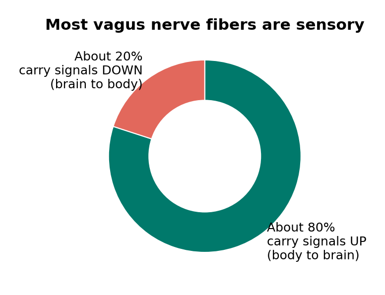
Source: classic animal fiber counts, as summarized by Prescott and Liberles 2022 and Bonaz et al. 2018. The split is approximate and varies between studies and species.
But notice the words "classic" and "in animals." Nobody has done a modern, complete fiber count of the human vagus nerve that settles the exact number. And one careful study suggests the split varies. In 1990, Prechtl and Powley counted every fiber in cross-sections of the abdominal vagus in rats using an electron microscope. They concluded that motor (efferent) fibers might make up over a quarter of the fibers there, a higher share than the widely accepted estimates. The precise share depends on the species, which branch of the nerve you count, and how you count.
What doesn't change is the big picture: most vagus traffic is sensory. The brain spends most of this cable on listening.
Hype Meter: "80% of your vagus nerve sends signals up to your brain"
2.6 The Route Up: From the Gut Wall to the Brainstem
Let's follow one afferent signal from the stomach to the brain. Imagine Sam's lunch stretching the wall of his stomach.
Step 1: The sensor. The ending of a vagal sensory fiber sits in the stomach wall. When the wall stretches, the ending fires electrical signals. Different vagal sensory neurons detect different things. In a 2016 study in mice, Williams and colleagues used genetic tools to show that one group of vagal sensory neurons responded to stretching of the stomach and intestine, while a different group responded to nutrients in the gut.
Step 2: The cell body. Sensory neurons have their cell bodies (the part that holds the nucleus) clustered in a lump outside the brain. The nodose ganglion is the cluster of cell bodies of vagal sensory neurons, located on the vagus nerve just below the skull in the neck; it is also called the inferior vagal ganglion. ("Nodose" means knotted, because it looks like a knot on the nerve.) Each nodose neuron has a long fiber reaching down to the organ and another fiber reaching up into the brainstem, so a signal passes straight through on its way up. A smaller group of vagal sensory neurons sits in a second cluster just above it, the jugular ganglion. In mice the two are fused into one, but in humans they are separate.
Step 3: The first stop in the brain. The nucleus tractus solitarius (NTS), also called the nucleus of the solitary tract, is a cluster of neurons in the medulla of the brainstem that receives incoming sensory signals from the vagus nerve and passes them on to other brain areas. The Prescott and Liberles review says nodose neurons send their signals mainly to the NTS, and that vagal fibers release the neurotransmitter glutamate there to excite NTS neurons. (Much of this wiring was mapped in animals, but the general layout is well established.)
From the NTS, information spreads to brain areas that control breathing and heart rate, hunger and stress, and conscious feeling. The NTS is like the city's central post office: it sorts the incoming mail and forwards it.
2.7 The Route Down: The Dorsal Motor Nucleus and the Vago-Vagal Reflex
Now follow the traffic the other way. The dorsal motor nucleus of the vagus (DMV) is a cluster of neurons in the medulla, next to the NTS, whose fibers travel down the vagus nerve to carry efferent (motor) signals to the stomach and intestines. These fibers don't usually connect directly to the gut's muscle. Instead, they connect to neurons of the enteric nervous system, which then carry out the order. Remember the neighborhood council from Chapter 1? The DMV is city hall calling the council, not doing the street repairs itself.
Two nuclei, side by side
 NTS and DMV look like alphabet soup at first, but there's a pattern: the NTS is where my sensory fibers arrive, and the DMV is where my motor fibers depart. They sit right next to each other in the brainstem, so a message can come in and a reply can go out almost immediately.
Because the NTS and DMV are neighbors and are wired to each other, the brainstem can run a loop: a sensory signal comes up the vagus, and a motor command goes back down the same nerve. Scientists call this a vago-vagal reflex (the vagus talking to itself through the brainstem). A 2016 review by Travagli and Anselmi describes these brainstem circuits as connecting incoming vagal fibers, NTS neurons, and outgoing fibers from the DMV, and notes that they are highly changeable, even shifting after stress.
Here's a real example. When a meal stretches your stomach, a 2013 review by Farré and Tack explains, stretch-sensing receptors trigger the upper stomach to relax and make room, a response called gastric accommodation, partly through vago-vagal reflexes. Later, when nutrients reach the first part of the small intestine, signals traveling mainly through vago-vagal reflexes slow down how fast the stomach empties. Your stomach is like an expandable backpack, and the vagus helps unzip the extra pocket when the load arrives.
flowchart LR
    A[Meal stretches stomach wall] --> B[Vagal sensory endings fire]
    B --> C[Signal passes nodose ganglion]
    C --> D[NTS in brainstem]
    D --> E[DMV in brainstem]
    E --> F[Efferent signal down the vagus]
    F --> G[Enteric neurons relax upper stomach]
Notice what this loop does not need: your conscious mind. The brainstem handles it.
2.8 The Other Wires: Spinal Afferent Nerves
The vagus is not the gut-brain connection. A 2013 review by Brookes and colleagues describes gut sensory neurons as organized into three separate pathways to the central nervous system: the vagal pathway, plus two pathways that enter the spinal cord, one in the middle of the back (thoracolumbar) and one lower down (lumbosacral).
A spinal afferent nerve is a sensory nerve fiber that carries signals from an organ, such as the gut, to the spinal cord rather than directly to the brainstem, with its cell body in a ganglion beside the spinal cord. From the spinal cord, signals travel up to the brain. A 2018 review by Brierley, Hibberd, and Spencer explains that spinal afferents reaching the colon and rectum come in many types that can detect gut contents, gentle and strong stretching, and chemicals released during inflammation. They give rise to sensations of discomfort, bloating, urgency, and pain.
Here's how the two wiring systems compare in general terms:
| Vagal afferents | Spinal afferents | |
|---|---|---|
| Path to the brain | Directly to the brainstem (NTS) | Into the spinal cord, then up to the brain |
| Cell bodies | Nodose and jugular ganglia, in the neck | Ganglia beside the spinal cord |
| Typical messages | Stretch, nutrients, gut hormones, some inflammation signals; fullness and nausea | Stretch, inflammation, injury; discomfort, urgency, and much of gut pain |
| Gut coverage | Most of the digestive tract | Whole gut, including the end of the colon and rectum |
When someone has gut pain, much of that message can travel through spinal routes, which is one reason cutting or stimulating the vagus doesn't simply switch gut feelings on or off. Chapter 8 explores pain pathways in detail.
2.9 Interoception: Sensing the Inside of Your Body
You have senses that point outward: sight, hearing, touch, taste, smell. You also have senses that point inward. You can tell when you are hungry, full, thirsty, out of breath, or when your heart is pounding. Scientists have a word for this.
Interoception is the process by which the nervous system senses, interprets, and combines signals from inside the body, such as fullness, nausea, heartbeat, and breathing, creating a moment-to-moment map of the body's internal state. A 2018 expert roadmap led by Sahib Khalsa describes interoception as working at both conscious and unconscious levels, and notes that problems with interoception are increasingly recognized in conditions such as anxiety disorders, mood disorders, and eating disorders. That doesn't mean interoception causes those conditions; researchers are still working out how the pieces fit.
Vagal and spinal afferents are the raw data for gut interoception. Much of it never reaches awareness, but some does, like Sam's tight stomach at the start line. The 2013 Brookes review notes that gut sensory neurons give rise to sensations such as fullness, bloating, nausea, discomfort, urgency, and pain.
Riya knows one of those sensations well. During her Crohn's flares, she often feels nauseated, even when she hasn't eaten anything unusual. (Riya is a fictional 16-year-old with Crohn's disease who appears throughout this book.) At one visit she asks her gastroenterologist, Dr. Alvarez, why inflammation in her intestine would make her feel sick to her stomach. Dr. Alvarez explains that the inflamed gut releases chemicals that can switch on sensory nerve endings. Reviews of animal research describe some gut sensory fibers that stay quiet until inflammatory chemicals turn them on. Some of these signals travel up the vagus to the NTS, which sits among the brainstem circuits that a 2014 review by Babic and Browning describes as central to nausea and vomiting. "Your gut isn't imagining it, and neither are you," Dr. Alvarez says. "The nausea is real information traveling a real pathway."
If you're going through this
If your gut condition makes you feel nauseated, full, or uncomfortable at unpredictable times, those feelings are real signals traveling real nerves, not something you're making up. It's worth keeping track of when they happen and telling your care team, because nausea and early fullness can point to things they can treat. You don't have to "push through" a meal or a class just to prove you're fine; it's okay to ask your school nurse or counselor for a plan.
2.10 Sensor Cells in the Gut Lining
The nerve endings in the gut wall don't sit right at the surface where food touches. Between them and your lunch is a single layer of lining cells. So how do nerves find out what's in the gut? Part of the answer is a special group of sensor cells scattered through that lining.
Think of them as smoke detectors spaced along a hallway, each sampling the air and sounding an alarm when it finds something.
An enteroendocrine cell is a hormone-releasing sensor cell scattered among the lining cells of the stomach and intestines, which detects nutrients and other substances in the gut and responds by releasing hormones and other signals. ("Entero" means intestine and "endocrine" means hormone-releasing.) They are rare: the Prescott and Liberles review says they make up about 1% of intestinal lining cells. But because the gut lining is enormous, that still adds up to a lot of sensors.
What do they release? A gut hormone is a chemical messenger made by cells of the digestive tract, mostly enteroendocrine cells, that travels through the blood or acts on nearby nerves to influence digestion, appetite, and blood sugar. A 2019 review by Gribble and Reimann describes gut hormones as made by scattered cells along the intestinal lining that signal nutrient absorption, food intake, and blood sugar control. Some travel through the bloodstream; others act locally on the vagal sensory endings next door.
For decades, scientists thought enteroendocrine cells talked to nerves only this indirect way, by releasing hormones that drift over to nearby nerve endings. Then a lab at Duke University found something surprising. The lab had noticed that some enteroendocrine cells in mice have a long, foot-like extension, which they named a neuropod. In 2015, Bohórquez and colleagues reported that neuropods reach out and directly contact nerves.
In 2018, the same lab, with first author Melanie Kaelberer, published a study in Science showing that in mice, these cells form actual synapses with vagal sensory neurons and pass signals to them using glutamate, a fast neurotransmitter. A neuropod cell is an enteroendocrine cell that forms synapse-like connections with nerve fibers and sends signals to them quickly, so far described mostly in mice. The team reported that in lab dishes where these gut cells were grown together with vagal neurons, connected neurons responded within milliseconds. A 2022 follow-up study by Buchanan and colleagues found that in mice, neuropod cells help the animals tell real sugar from artificial sweetener, and the preference for sugar depended on the cells' glutamate signaling.
Let's apply the six questions from Chapter 1, Section 1.13 to the 2018 study.
How to read a gut-brain claim: the 2018 neuropod cell study
| Question | This study |
|---|---|
| Mouse or human? | Mouse. The experiments used genetically modified mice, mouse gut tissue grown in dishes (organoids), and mouse vagal neurons. No human tissue was tested for synapses. |
| Correlation or cause? | Mostly cause, in mice. The team switched the gut cells on and off with light (optogenetics) and blocked glutamate receptors, then watched vagal nerve activity change. That tests cause, but only in mice. |
| How many? | Small groups, which is normal for this kind of lab work: for example, 5 to 9 mice for some nerve recordings and 9 connected cell pairs for the timing measurement. |
| How long? | Minutes to hours. These were short experiments on nerve signals, not long-term studies of health or behavior. |
| Who paid for it? | The paper lists NIH grants, the AGA-Elsevier pilot research award, DARPA, The Hartwell Foundation, The Dana Foundation, and The Grass Foundation; one author was supported as a Howard Hughes Medical Institute fellow. |
| Is something being sold? | No product, but the paper states that some findings were used to file a provisional patent application. |
The conclusion: this is strong, careful basic science showing a new kind of gut-to-nerve connection in mice. It does not show that humans have the same synapses working the same way, and it says nothing yet about treating any disease.
Hype Meter: "Your gut cells are wired straight to your brain"
2.11 Feeling Full: CCK, GLP-1, and Satiety Signals
Back to Sam at lunch on the day of his chemistry final. After a few bites, he feels full. What's actually telling his brain to stop eating?
A satiety signal is any signal from the gut or body that tells the brain that enough food has been eaten, producing the feeling of fullness and reducing the urge to keep eating. Satiety signals come in two main flavors: nerve signals from stretch (the stomach is physically filling up) and chemical signals from the gut hormones released when nutrients arrive.
Cholecystokinin (CCK) is a gut hormone released by enteroendocrine cells in the small intestine (called I-cells), mainly when protein-rich and fat-rich food arrives, that helps digest food and acts as a satiety signal partly by exciting vagal sensory nerves. CCK was discovered in 1928 as a substance that makes the gallbladder contract, which is where its name comes from. A 2017 review by Jens Rehfeld lists its jobs: squeezing the gallbladder, boosting digestive enzymes from the pancreas, changing gut movement, reducing stomach acid, and producing satiety. In 1973, Gibbs, Young, and Smith reported that injecting CCK reduced how much food rats ate.
Smith and Gibbs later recalled that CCK was simply the first gut hormone their team chose to test, and the first experiments worked. Human studies have since backed up the basic idea: a 2023 review of 15 randomized trials in healthy adults, by Warrilow and colleagues, found that CCK helps bring a meal to an end sooner (satiation), but found no evidence that it causes long-term weight loss. The 2018 Bonaz review describes CCK, GLP-1, and another gut hormone called PYY as reaching the brain through vagal afferents, though much of the evidence for that vagal route comes from animals.
GLP-1 (glucagon-like peptide 1) is a gut hormone released after eating by enteroendocrine cells called L-cells in the intestinal lining, that boosts insulin release when blood sugar is high, slows stomach emptying, and reduces food intake. A 2019 review by Müller and colleagues lists those three effects. GLP-1 has become famous outside biology class, too. The Gribble and Reimann review notes that drugs that copy GLP-1 (GLP-1 mimetics) are licensed medicines, used for type 2 diabetes and for obesity. As of 2026, some of these drugs are FDA approved for obesity in teens 12 and older. For example, semaglutide, sold as Wegovy, gained that approval in December 2022. Those medicines are prescribed and monitored by doctors; this chapter teaches how the natural hormone works, not who should take them.
| Signal | Where it comes from | What triggers it | Main effects | Route to the brain |
|---|---|---|---|---|
| Stomach stretch | Stretch-sensing vagal endings in the stomach wall | Food volume | Fullness; triggers gastric accommodation | Vagal afferents to NTS |
| CCK | Enteroendocrine cells (I-cells) in the small intestine | Protein-rich and fat-rich food | Gallbladder squeezes; fullness; slower stomach emptying | Mainly vagal afferents (mostly animal evidence) |
| GLP-1 | Enteroendocrine cells (L-cells) in the intestinal lining | A meal | More insulin when blood sugar is high; slower stomach emptying; less eating | Vagal afferents and the bloodstream |
So what happened to Sam? Stress may change how his stomach and its brainstem circuits behave (the Travagli review notes these circuits shift after stress), and the sympathetic system slows digestion when he's keyed up. His IBS may also make normal stretch feel bigger (Chapter 8 explains how). A test-day sandwich can feel huge when the stomach isn't relaxing to make room.
2.12 Trying It Yourself: The Vagus Signal Router
You now know all the parts: afferent fibers carrying news up, the nodose ganglion, the NTS and DMV in the brainstem, efferent fibers carrying commands down, the spinal route, and gut hormones like CCK. The simulation below puts them together. Teal pulses are afferent signals, and coral pulses are efferent signals.
Diagram: Vagus Signal Router
Open the Vagus Signal Router fullscreen
MicroSim specification: Vagus Signal Router
Type: microsim
sim-id: ch02-vagus-signal-router
Library: p5.js
Status: Implemented
Bloom Level: Analyze
Bloom Verb: compare
Learning Objective: The learner will compare afferent (gut-to-brain) and efferent (brain-to-gut) vagus signaling across four everyday events, and contrast the vagal route with the spinal route.
Canvas: 540 px drawing area plus 120 px controls (iframe 662 px); responsive to about 380 px wide, where the info panel moves below the diagram.
Controls: Start/Pause, Reset, "Cut the vagus (vagotomy)" checkbox, and four event buttons: Meal stretches stomach, Fat arrives (CCK), Gut inflammation, Stressful thought.
Rules: Meal: 4 afferent stretch pulses up the vagus; each arrival at the NTS sends an efferent "relax" pulse down (gastric accommodation). Fat: an enteroendocrine cell flashes "CCK," then 4 afferent pulses rise (satiety signal); some arrivals send an efferent "empty more slowly" pulse. Inflammation: 3 afferent pulses up the vagus and 3 up the spinal route (nausea and unwellness via the vagus; discomfort and pain via spinal nerves); some vagal arrivals send an efferent anti-inflammatory pulse, labeled as studied mostly in animals. Stressful thought: starts in the brain; DMV output dims and 3 sympathetic pulses travel down the spinal route (digestion slows). With the vagus cut, vagal pulses stop at the cut in both directions; spinal pulses still pass.
Feedback: Counters for signals up and signals down; a status panel names what the gut and brain are doing; a fixed fiber bundle shows 8 of 10 fibers sensory (classic animal counts, about 80%).
Provenance: Sections 2.5 to 2.11 (Prescott and Liberles 2022; Bonaz 2018; Travagli and Anselmi 2016; Farré and Tack 2013; Brookes 2013; Brierley 2018). Pulse speeds and counts are illustrative.
Start with Meal stretches stomach and watch the order of events: teal pulses rise to the NTS first, and only then do coral pulses leave the DMV and travel down. That's the vago-vagal reflex: information up, a command down. Watch the counters, too. After a few events, notice how many more signals travel up than down, and compare that with the 8-of-10 fiber bundle.
Then try Gut inflammation and notice that two routes light up: the vagus and the spinal nerves. Now check Cut the vagus (vagotomy) and repeat each event. Predict first: which events still reach the brain? Stretch and CCK signals stop at the cut, and the stomach never gets its "relax" command. But inflammation still sends signals up the spinal route, and a stressful thought still slows digestion through sympathetic nerves. Keep that result in mind for the At the Bedside case.
What this model leaves out
- A real vagus nerve has tens of thousands of fibers in each branch, not ten, and the 8-of-10 split is an approximate figure from animal counts.
- Real signals are streams of electrical impulses of different speeds; gut hormones like GLP-1 also travel through the blood, which the sim doesn't show.
- The brainstem connects to many higher brain areas (hunger, stress, emotion); the sim shows only the NTS and DMV.
- The sim doesn't show the heart, lungs, and other organs the vagus also serves, or the pelvic nerves to the end of the colon.
- Timings are made up for teaching, and real vagotomy effects on symptoms vary from person to person.
2.13 Vagal Tone: Can You Measure Your Vagus Nerve?
Wellness posts love the phrase "vagal tone." Here's what it means.
Vagal tone is the ongoing, background level of activity in the vagus nerve's efferent (motor) fibers, most often estimated through its calming effect on the heart. Even at rest, the vagus gently brakes your heart.
Researchers usually estimate vagal tone indirectly, using heart rate variability (HRV), the small, normal changes in time between one heartbeat and the next. A 2017 review by Laborde, Mosley, and Thayer explains that HRV can index cardiac vagal tone, meaning the parasympathetic contribution to controlling the heart. But the same review warns that HRV findings are easy to misinterpret and need careful methods. Breathing rate, posture, caffeine, exercise, time of day, and the exact way the measurement is taken all change HRV.
The 2018 Bonaz review notes that lower vagal tone has been described in some people with IBD and IBS. That's an interesting association, but it doesn't tell us which comes first: low vagal tone could contribute to gut problems, gut problems could lower vagal tone, or a third factor (like stress, poor sleep, or medicines) could affect both.
Your watch measures your heart, not your gut
 A smartwatch HRV number is an indirect estimate of my effect on your heart, and it shifts with breathing, sleep, caffeine, and how still you sit. It doesn't directly measure my gut fibers, and it can't tell you whether your vagus nerve is "healthy."
Posts promise that splashing cold water on your face, taking an ice bath, humming, singing, or gargling will "reset" or "activate" your vagus nerve. Some of this has a grain of truth. In a 2018 lab study of 61 healthy adults, Jungmann and colleagues found that briefly cooling the side of the neck slightly raised HRV and lowered heart rate compared with a no-cooling condition; the authors called it a first step toward designing a wearable device. A 2018 systematic review by Zaccaro and colleagues, which included 15 studies of healthy people, found that slow breathing (under 10 breaths a minute) increased HRV and was linked to feeling calmer.
But there's a big leap from "a short change in heart-rate measurements in healthy volunteers" to "cures anxiety" or "heals your gut."
Hype Meter: "Cold plunges, humming, or gargling reset your vagus nerve and cure anxiety"
For families
If your teen gets stomachaches or loses their appetite before tests, games, or performances, the feeling is real: stress signals really do change how the stomach moves and how gut signals reach the brain. Saying "it's all in your head" can feel dismissive, even though the brain is involved. Try something like, "Your body is reacting to a big day; what would help right now?" If symptoms happen often, interfere with eating, or come with weight loss, pain that wakes them up, or blood in the stool, that's a reason to see a doctor, not to try a vagus "hack."
2.14 When Surgeons Cut the Vagus: Vagotomy
In the mid-1900s, ulcers (open sores in the lining of the stomach or the first part of the small intestine) often needed surgery. Doctors knew stomach acid played a big part, and that the vagus helps tell the stomach to make acid. So surgeons tried cutting it.
Vagotomy is a surgery that cuts all or part of the vagus nerve to the stomach, historically done to reduce stomach acid in people with ulcers. The American surgeon Lester Dragstedt at the University of Chicago pioneered vagotomy for peptic ulcer disease in the 1940s, according to a 1994 history by Modlin and Darr.
Cutting the main trunks (truncal vagotomy) reduced acid, but it also removed the vagus's efferent control of stomach movement. A clinical overview for doctors on Patient.info explains that truncal vagotomy alone could stop the stomach from emptying properly, so surgeons added a second operation to widen the stomach's exit (a pyloroplasty). That overview also notes that diarrhea is common after vagotomy. Surgeons developed more selective versions that cut only the branches to the acid-making part of the stomach, which had fewer side effects but higher rates of ulcers coming back.
Then the reasons for the surgery largely disappeared. Medicines that powerfully reduce stomach acid became available, and doctors discovered that a bacterium, Helicobacter pylori, causes many ulcers and can be treated with antibiotics. A 2014 review by Lagoo, Pappas, and Perez concludes that the role of vagotomy in ulcer care is now significantly limited, kept mainly for emergencies and rare hard-to-treat cases.
Doctors still meet older patients who had a vagotomy decades ago, like the patient below. And interest in the vagus has turned in a new direction: instead of cutting it, researchers stimulate it. Implanted vagus nerve stimulators are FDA approved for some people with hard-to-treat epilepsy and depression. In August 2021 the FDA approved one called Vivistim, used during rehabilitation therapy, for people with long-lasting, moderate to severe arm weakness after an ischemic stroke. And in July 2025 the FDA approved one called the SetPoint System for adults with moderate to severe rheumatoid arthritis who hadn't responded well to biologic or targeted drugs. Vagus nerve stimulation is not approved for Crohn's disease, IBS, or anxiety.
One related device is worth knowing about, so you don't mix it up: in June 2019 the FDA authorized IB-Stim, a small stimulator worn behind the ear that sends mild pulses to nerves in and around the ear, as an add-on to help reduce IBS-related belly pain in patients 11 to 18 years old. That was a De Novo authorization, the FDA route for new kinds of lower-risk devices. It is a different, lighter review than the full premarket approval that implanted vagus nerve stimulators go through, and it does not mean implanted vagus nerve stimulation is approved for IBS. Chapter 17 covers these devices in detail.
At the Bedside: Full After a Few Bites
Meet the patient (fictional)
Walter is 74 and recently moved to be closer to his daughter. At his first visit with a new gastroenterologist, he explains that he has felt full after just a few bites for most of his adult life, and that he has bouts of loose, urgent stool, especially after larger meals. Lately he has started skipping family dinners at restaurants because he worries about needing a bathroom quickly. His daughter mentions that he "had stomach surgery back in the 1970s."
What the doctor checks: The doctor takes a careful history, including old surgical records. They show that Walter had a truncal vagotomy and pyloroplasty for a duodenal ulcer in 1976. The doctor asks about weight changes, what his meals look like, when symptoms happen, and any warning signs such as blood in the stool, vomiting, or night sweats. Because Walter is older and his symptoms are long-standing, the doctor also reviews his medicines and orders tests to make sure nothing new (such as an infection or another bowel disease) is adding to the problem.
Connecting the signals: When the main vagal trunks were cut, Walter lost most of the efferent signals from his DMV to his stomach. Normally, a meal stretches the stomach, afferent signals reach the NTS, and efferent signals come back down to relax the upper stomach (gastric accommodation). Without that command, his stomach can't expand as easily, so it feels full quickly. The widened stomach exit from his pyloroplasty also lets food leave faster, and diarrhea is a known, common effect after vagotomy.
Diagnosis: Long-term symptoms after vagotomy (sometimes called post-vagotomy syndrome): early fullness and diarrhea, with tests showing no new disease.
Why it happens: This is the Vagus Signal Router's "cut" setting in real life. Signals up and down the vagus can't cross the cut, so the brainstem can't run its vago-vagal reflexes for the stomach. Walter's enteric nervous system still works (it runs local reflexes, as Chapter 1 showed), and his spinal nerves still carry sensations, so his gut isn't silent, just missing an important line of communication.
The mind-body side: Walter's biggest problem right now isn't the biology; it's that he has started avoiding meals with his family. Worry about urgent bathroom trips can make people feel more on edge during meals, and stress signals can make the gut more reactive, which feeds the worry. The care team takes this seriously: they ask how symptoms affect his life, and they may suggest planning ahead (for example, knowing where restrooms are) and talking with a counselor if the worry keeps growing.
Treatment today: There's no way to reconnect a vagus nerve cut decades ago. Care focuses on symptoms. A dietitian can help Walter find a meal pattern that works for him, and his doctor may consider medicines that reduce diarrhea, depending on the cause and his other health conditions. Each plan is individual and is worked out with his care team.
What's new in research: Researchers are now testing whether stimulating the vagus could calm gut inflammation. In a 2016 pilot study, Bonaz and colleagues implanted vagus nerve stimulators in 7 adults with active Crohn's disease and followed them for 6 months. Five improved, with lower inflammation markers, while two were removed from the study at 3 months because they got worse. There was no comparison group. A 2023 proof-of-concept study by Sahn and colleagues tried a noninvasive stimulator clipped to the ear in 22 young people aged 10 to 21 with mild to moderate IBD for 16 weeks; a stool marker of gut inflammation fell by at least half in 11 of the 17 participants who started with high levels. In the first 4 weeks, each participant got 2 weeks of real stimulation and 2 weeks of a sham (fake) version; after that, everyone got real stimulation, so most of the study had no control group. Evidence grade: Preliminary. Vagus nerve stimulation is not approved for Crohn's disease.
Your turn: reason through a variation
Suppose Walter had instead had a highly selective vagotomy, which cuts only the vagal branches to the acid-making part of the stomach and leaves the branches to the stomach's lower part and the intestines. Would you expect his symptoms to be the same, milder, or worse? What trade-off did surgeons accept with this version?
Answer: Milder. Because more efferent control of stomach emptying is kept, the stomach can still empty in a more controlled way without a pyloroplasty, so diarrhea is less of a problem. The trade-off is that cutting fewer fibers reduces acid less completely, so ulcers came back more often after highly selective vagotomy.
This is a fictional teaching case. It is not medical advice. If you or someone you know has symptoms like these, talk to a doctor.
Common Misconceptions
Myth: The vagus nerve mostly sends commands from the brain down to the gut.
Reality: Most vagus fibers are sensory. Classic fiber counts in animals put about 80% of them as afferent, carrying information from the body up to the brainstem.
Myth: The vagus nerve is just a "calm down" switch.
Reality: The vagus carries a huge range of information (stretch, nutrients, hormones, inflammation) and helps run reflexes such as gastric accommodation. Its parasympathetic effects matter, but it is mainly a sensory cable, not a relaxation button.
Myth: Your smartwatch measures your vagal tone directly.
Reality: HRV is an indirect estimate of the vagus nerve's effect on the heart, and it changes with breathing, posture, caffeine, sleep, and how it's measured. It doesn't measure the vagus fibers to the gut.
Myth: The vagus is the only way the gut talks to the brain.
Reality: Spinal afferent nerves carry many gut sensations, including much of gut pain, and gut hormones and immune signals travel through the blood. The vagus is one important route among several.
Myth: Neuropod cells prove your gut is wired directly into your brain.
Reality: Neuropod cells were described in mice, where they connect to vagal sensory neurons, which then signal the brainstem. Whether human gut cells work the same way is still being studied.
Your Gut Check
Test yourself. Try each one before you open the answer.
1. Claim Check: "Hum for 5 minutes a day to activate your vagus nerve and heal your gut. Your gut-brain connection will thank you!"
Grade: Hype. Slow breathing can raise heart rate variability in healthy people, and humming may slow breathing, but HRV is an indirect estimate of the vagus nerve's effect on the heart, not the gut. None of the evidence in this chapter shows that humming changes gut function or heals any gut condition, and "activate" is vague. A careful version would say: "Slow breathing can briefly change heart-rate measures linked to the vagus nerve, and some people find it calming; it hasn't been shown to treat gut disease."
2. Predict: Sam is nervous before a race and eats a big bowl of pasta an hour before the start. Predict how his stomach might respond, using the parasympathetic and sympathetic branches.
Race nerves boost sympathetic (fight-or-flight) activity and dampen vagal output, so his stomach may relax and empty less smoothly, and digestion slows. The pasta stretches his stomach, and stretch signals still travel up the vagus, so he may feel uncomfortably full or queasy. The likely result is a heavy, unsettled stomach at the start line. This is a reason many athletes experiment with timing meals, ideally with advice from a coach or doctor.
3. Explain: Riya feels nauseated during Crohn's flares even when she hasn't eaten anything new. Explain the pathway from her inflamed intestine to the feeling of nausea.
Inflammation releases chemicals that can switch on sensory nerve endings in the gut wall. Afferent signals travel up the vagus nerve, past the nodose ganglion, to the NTS in the brainstem, which sits among the circuits that produce nausea; spinal afferents also carry discomfort signals. The brain turns this incoming information into the conscious feeling of nausea, which is an example of interoception. The feeling is real information from her gut, not something imagined.
4. Apply: In an experiment, rats are given an injection of CCK just before a meal. A second group gets the same injection after their vagus nerves have been cut. Predict the results.
The first group should eat less, because CCK is a satiety signal (Gibbs and colleagues showed this in rats in 1973). In the second group, the effect should be much weaker, because CCK works largely by exciting vagal sensory nerves, and the cut prevents those signals from reaching the NTS. If the vagotomized rats still ate a little less, that would suggest CCK also acts through other routes.
5. Read the data. Here are two estimates of how vagus nerve fibers split between sensory (afferent) and motor (efferent). The first is the classic figure as cited by Prescott and Liberles (2022); the second is from a careful rat study (Prechtl and Powley, 1990).
| Source | Animal and nerve region | Finding |
|---|---|---|
| Classic animal fiber counts: Foley and DuBois, 1937 (as cited by Prescott and Liberles, 2022) | Cat vagus nerve | About 80% of fibers sensory |
| Prechtl and Powley, 1990 | Rat abdominal vagus (6 animals examined in detail) | Motor fibers may be over a quarter of all fibers; 22,272 fibers counted in the main trunks, 26,930 summed across five branches |
5. What does this data show, and what can't it show?
Both sources agree that most vagus fibers are sensory, but they disagree on how many: if motor fibers are over 25%, sensory fibers are under 75%, not 80%. The branch total being about 21% higher than the trunk total shows that counting depends on where and how you count (the authors think extra fibers joined from outside the vagus). The data can't tell us the exact human figure, because both come from animals and the 1937 count is old and not easy to check. A useful follow-up would be a modern fiber count of human vagus nerves at several levels, using the same method for every branch.
Exercises
Remember
- What is the difference between an afferent signal and an efferent signal?
Answer
An afferent signal travels toward the central nervous system (usually sensory information), and an efferent signal travels away from it to muscles, glands, or organs (usually a command).
- Name the two brainstem nuclei of the vagus nerve covered in this chapter and say what each does.
Answer
The nucleus tractus solitarius (NTS) receives incoming sensory signals from the vagus. The dorsal motor nucleus (DMV) sends efferent signals down the vagus to the stomach and intestines.
Understand
- Explain why the statement "the vagus nerve is the brain's control cable for the gut" is misleading.
Answer
Most vagus fibers (about 80% by classic animal counts) are sensory and carry information up to the brain, so the nerve is mainly a listening line. Its efferent fibers also work through the enteric nervous system rather than controlling gut muscle directly.
- Explain why a smartwatch's HRV number is an indirect measure of vagal tone.
Answer
HRV reflects the vagus nerve's braking effect on the heart, mixed with many other influences such as breathing, posture, caffeine, and sleep. It doesn't record vagus nerve activity directly, and it says nothing direct about the vagus fibers to the gut.
Apply
-
Use the Vagus Signal Router. Trigger each of the four events three times with the vagus intact, and record the "Signals up" and "Signals down" counters. Then reset, cut the vagus, and repeat. Make a table of your results and describe which events still reach the brain after vagotomy, and by which route.
-
A friend says, "Gut hormones are just like nerve signals." Using CCK and GLP-1 as examples, describe one way gut hormones are like nerve signals and one way they are different.
Analyze
-
Compare the 2016 Williams mouse study (different vagal neurons sense stretch and nutrients) with the 2016 Bonaz pilot study of vagus nerve stimulation in 7 people with Crohn's disease. For each, answer three of the six questions (mouse or human, correlation or cause, how many). Which study tells you more about humans, and which is better at showing cause?
-
Sam feels sick before races and full early on test days. List at least three different pathways from this chapter (nerves, brainstem circuits, hormones, autonomic branches) that could contribute, and explain how you might design an observation or experiment to tell them apart.
Evaluate
-
A company sells a $200 earbud device that it says "stimulates your vagus nerve to fix your digestion." Using the evidence in this chapter, including what is and isn't FDA approved, judge this claim. What evidence would you need to see before you believed it?
-
Some writers say the "80%" figure should be dropped from textbooks because it comes from old animal counts. Others say it's still a useful approximation. Write a one-paragraph argument for which side you agree with, using the Prechtl and Powley data.
Create
-
Design an experiment, using mice, that would test whether neuropod cells are needed for the feeling of fullness after a sugary meal. Describe your groups, what you would change, what you would measure, and the result that would support your hypothesis.
-
Create a one-page infographic or comic strip that follows a single bite of food from the moment it stretches the stomach to the moment the stomach relaxes, labeling every afferent and efferent step and naming the nodose ganglion, NTS, and DMV.
Key Takeaways
- The vagus nerve (cranial nerve X) is the longest cranial nerve and the main parasympathetic nerve to the gut. It carries both afferent (gut-to-brain) and efferent (brain-to-gut) fibers.
- Most vagus fibers are sensory. Classic fiber counts in animals put the figure at about 80%, though careful counts show the exact share varies.
- Sensory signals pass through the nodose ganglion to the nucleus tractus solitarius in the brainstem; the dorsal motor nucleus next door sends commands back down, creating vago-vagal reflexes such as gastric accommodation.
- The vagus isn't the only route: spinal afferent nerves carry many gut sensations, including much of gut pain, and gut hormones also travel through the blood.
- Enteroendocrine cells sense nutrients and release gut hormones such as CCK and GLP-1, which act as satiety signals; in mice, some of these cells (neuropod cells) form fast synapses with vagal neurons.
- Vagal tone is usually estimated indirectly through heart rate variability. Claims that cold plunges, humming, or gargling "reset" the vagus nerve and cure anxiety are Hype.
- Vagotomy, once a common ulcer surgery, shows what the vagus does for the stomach: without it, accommodation and controlled emptying suffer.
You rode the whole highway!
 You can now trace a signal from the gut wall to the brainstem and back, and you know why most of my traffic heads uphill. Next, in Chapter 3, we'll meet the trillions of microbes living in your gut and find out what they might add to the conversation.